Sds page protocol for protein pdf Nueva Plymouth
SDS-PAGE for protein electrophoresis 2-D electrophoresis results. The 2-D protocols described herein are performed using Amersham Biosciences products. Equipment choices are discussed on page 12 and illustrated in Table 1. Introduction to two-dimensional (2-D) electrophoresis Two-dimensional electrophoresis (2-D electrophoresis) is a powerful and widely used
Title SDS-PAGE SOP Biomanufacturing
Native SDS-PAGE High Resolution Electrophoretic. SDS/PAGE MINI PROTEIN GEL Polyacrylamide gel electrophoresis (PAGE) is a widely used technique for separating proteins. The most widely used method was developed by Laemmli (Nature 227: 690-685, 1970) using the denaturing (SDS) discontinuous method. This protocol relies on the presence, Introduction to SDS-PAGE. This material is accompanied by a presentation on protein structure and principles behind denaturing samples and discontinuous gel electrophoresis.. The separation of macromolecules in an electric field is called electrophoresis.A very common method for separating proteins by electrophoresis uses a discontinuous polyacrylamide gel as a support ….
interaction permits boiling the protein complex in SDS sample buffer prior to SDS-PAGE analysis. Additional Resources for the HaloTag®Technology Technical Bulletins and Manuals TM260 HaloTag®Technology: Focus on Imaging Technical Manual Promega Publications Efficient high-throughput protein purification using the Magne™ HaloTag®Beads SDS-PAGE for protein electrophoresis Aim This section is taked and modified from the protocol provided by Kristian Dreij, Hanna Karlsson in SDS-PAGEprotocolbyAssay-protocol.com. The protocol written by Kristian Dreij, Hanna Karlsson in the course "Applications of Methods in
UIC REERENCE Pub No MAN0007891 Rev C.0 NuPAGE™ Bis-Tris Mini Gels Choosing a well format Thicker 1.5 mm gels with fewer wells are recommended for large samples (>30 μL). Thinner 1 mm gels are recommended for blotting because of better protein transfer. Download SDS-PAGE protocol as a PDF . SDS-PAGE, with full name of sodium dodecyl sulfate polyacrylamide gel electrophores, is the most widely used technique to separate proteins from complicated samples of mixture, plays key roles in molecular biology and wide range of subfield of biological research. Being present a electricity, proteins migerate towards the negative …
Recommended SDS PAGE Stain Protocols Kits like GelCode Blue from Pierce and Biosafe Coomassie from Biorad are NOT compatible for in-gel digestion and mass spectrometry analysis unless you do a fixing step first. Oct 20, 2018 · SDS-PAGE (Polyacrylamide Gel Electrophoresis), is an analytical method used to separate components of a protein mixture based on their size. The technique is based upon the principle that a charged molecule will migrate in an electric field …
Oct 20, 2018 · The method and detailed procedure of SDS-PAGE for silk proteins are exactly the same as for other proteins, but the electrophoresis profile of silk protein is often unsatisfactory. The main reason is that their molecular masses are too large, and the regenerated liquid silk is easily coagulated and denatured, resulting in a significant adverse effect on normal … pdf. PROTOCOL Tricine–SDS-PAGE. Hoi Suparat. Here I describe a protocol for Tricine–SDS-PAGE, which includes efficient methods for Coomassie blue or silver staining and electroblotting, thereby increasing the versatility of the approach. and generates SDS-protein complexes that are mostly characterized by a uniform charge-to-mass
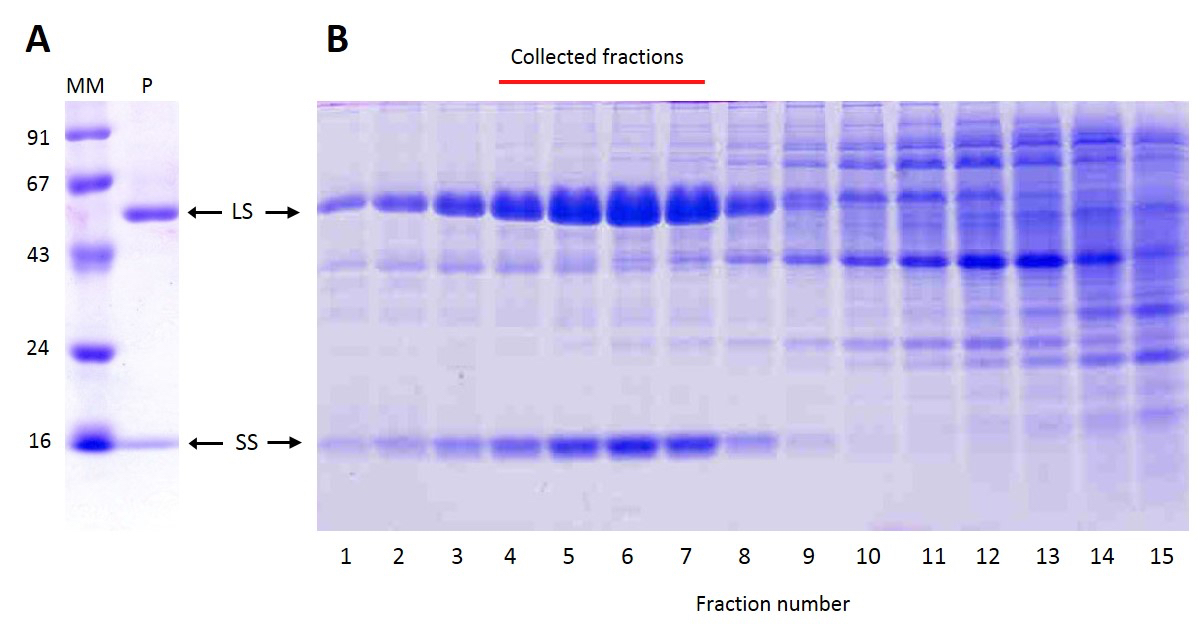
Aug 29, 2016 · PDF SDS-PAGE plays a key role in the study of protein based variation among different brassica species. The present study aimed to develop an efficient Sodium Dodecyl Sulphate Poly Acrylamide Together, Laemmli–SDS-PAGE and Tricine–SDS-PAGE cover the protein mass range 1–500 kDa. Answering the following questions should help you to identify the optimal solution for a specific sep-aration problem and help you to decide whether this …
Introduction to SDS-PAGE. This material is accompanied by a presentation on protein structure and principles behind denaturing samples and discontinuous gel electrophoresis.. The separation of macromolecules in an electric field is called electrophoresis.A very common method for separating proteins by electrophoresis uses a discontinuous polyacrylamide gel as a support … Protein gel electrophoresis technical handbook Electrophoresis run conditions 2 For ordering information refer to page XX. For quick reference on the protocol please refer to page XX. 3 ntse Cnto Electrophoresis overview 4 Select precast gel Gel selection guide 8 Gels 10 The resultant SDS-protein complexes are highly
Native SDS-PAGE of LLC-PK 1 cell proteome and model metalloproteins. Neither SDS-PAGE nor BN-PAGE was able to accomplish the goal of acceptable protein separation with retention of enzymatic activity. A new method or effective changes to these protocols were needed to achieve both. We chose to modify SDS-PAGE. Bio 6 – SDS-PAGE Lab Objectives Upon completion of this laboratory you will understand how to load and run protein samples on an SDS-polyacrylamide gel, stain the gel, and analyze the resulting bands of protein on the gel to estimate the molecular weight of each protein.
Sodium dodecyl sulfate (SDS)-polyacrylamide gel electrophoresis (PAGE) is an analytical method that enables protein separation based on their molecular mass. SDS is an anionic detergent, which facilitates the denaturation of the native proteins by disturbing the non-covalent forces. Together, Laemmli–SDS-PAGE and Tricine–SDS-PAGE cover the protein mass range 1–500 kDa. Answering the following questions should help you to identify the optimal solution for a specific sep-aration problem and help you to decide whether this …
— If protein phosphorylation is to be studied, include phosphatase inhibitors such as okadaic acid, calyculin A, and vanadate. When working with a new sample, use at least two different cell disruption protocols and compare the protein yield (by protein assay) and qualitative protein content (by SDS-PAGE) Peccoud Lab Protocol: visualizing proteins via sodium dodecyl sulfate-polyacrylamide electrophoresis (SDS-PAGE) Introduction. Because cellular extracts contain thousands of different proteins at a wide range of concentrations, it is often difficult to detect and measure specific proteins in these mixes, even when proteins are expressed at high concentrations.
Introduction to SDS-PAGE. This material is accompanied by a presentation on protein structure and principles behind denaturing samples and discontinuous gel electrophoresis.. The separation of macromolecules in an electric field is called electrophoresis.A very common method for separating proteins by electrophoresis uses a discontinuous polyacrylamide gel as a support … Use PAGE gels, a Bradford assay, a Lowry assay or a BCA assay. Bovine serum albumin (BSA) is a frequently-used protein standard. Preparation of samples for loading into gels. To denature, use a loading buffer with the anionic denaturing detergent sodium dodecyl sulfate (SDS), and boil the mixture at 95-100°C for 5 minutes. Electrophoresis. 1).
Chapter 14

Protein Gel Protocol.. Use PAGE gels, a Bradford assay, a Lowry assay or a BCA assay. Bovine serum albumin (BSA) is a frequently-used protein standard. Preparation of samples for loading into gels. To denature, use a loading buffer with the anionic denaturing detergent sodium dodecyl sulfate (SDS), and boil the mixture at 95-100°C for 5 minutes. Electrophoresis. 1)., SDS-PAGE Protocol Mutated from the SDS-PAGE protocol written by the Lord of the Flies Pouring the resolving gel 1. Clean glass plates with soap and water, then with ethanol. Assemble the glass plates and spacers. 2. For large gels, seal with 1 mL of resolving gel. Remove 2 mL and when.
SDS-PAGE and Western Blotting Protocol. SODIUM DODECYL SULFATE-POLYACRYLAMIDE GEL ELECTROPHORESIS (SDS-PAGE) All Hycult Biotech products are subject to strict quality control procedures., Aug 29, 2016 · PDF SDS-PAGE plays a key role in the study of protein based variation among different brassica species. The present study aimed to develop an efficient Sodium Dodecyl Sulphate Poly Acrylamide.
Bio 6 – SDS-PAGE Lab

Native SDS-PAGE High Resolution Electrophoretic. In order to identify proteins by size, protein standards of a known size are loaded along with samples and run under the same conditions. This video presents an introduction to SDS-PAGE by first explaining the theory behind it and later demonstrating its step-by-step procedure. https://fr.wikipedia.org/wiki/Bleu_de_Coomassie pdf. PROTOCOL Tricine–SDS-PAGE. Hoi Suparat. Here I describe a protocol for Tricine–SDS-PAGE, which includes efficient methods for Coomassie blue or silver staining and electroblotting, thereby increasing the versatility of the approach. and generates SDS-protein complexes that are mostly characterized by a uniform charge-to-mass.
August 18, 2003 Edition Page 2 Mini-Protean SDS-PAGE Protocol Casting the Gel 1] Assemble glass plates and spacers in gel casting apparatus–see BioRad instruction manual. 2] Mix the components for the resolving gel as described in the Mini-Protean II protocol. May 14, 2014 · Sds-Page 1. Sodium Dodecylsulfate Polyacrylamide Gel Electrophoresis(SDS-PAGE) BY: JANE LOH JENNY ONG 2. Overview a. Introduction b. Principle of Sds-page c. Gel preparation d. Process of Sds-page e. Visualization of protein f. Applications g. Advantages and disadvantages h. Conclusion i. References 3.
SDS/PAGE MINI PROTEIN GEL Polyacrylamide gel electrophoresis (PAGE) is a widely used technique for separating proteins. The most widely used method was developed by Laemmli (Nature 227: 690-685, 1970) using the denaturing (SDS) discontinuous method. This protocol relies on the presence Aug 29, 2016 · PDF SDS-PAGE plays a key role in the study of protein based variation among different brassica species. The present study aimed to develop an efficient Sodium Dodecyl Sulphate Poly Acrylamide
August 18, 2003 Edition Page 2 Mini-Protean SDS-PAGE Protocol Casting the Gel 1] Assemble glass plates and spacers in gel casting apparatus–see BioRad instruction manual. 2] Mix the components for the resolving gel as described in the Mini-Protean II protocol. In SDS- PAGE, the protein mixture is denatured by heating at 100 qC in the presence of excess SDS and a reducing reagent is employed to break disu lfide bonds. Under these conditions, all reduced polypeptide bind the same amount of SDS on a weight basis (1.4g SDS/g polypeptide) independent of the amino acid composition and sequence of the protein.
pdf. PROTOCOL Tricine–SDS-PAGE. Hoi Suparat. Here I describe a protocol for Tricine–SDS-PAGE, which includes efficient methods for Coomassie blue or silver staining and electroblotting, thereby increasing the versatility of the approach. and generates SDS-protein complexes that are mostly characterized by a uniform charge-to-mass Peccoud Lab Protocol: visualizing proteins via sodium dodecyl sulfate-polyacrylamide electrophoresis (SDS-PAGE) Introduction. Because cellular extracts contain thousands of different proteins at a wide range of concentrations, it is often difficult to detect and measure specific proteins in these mixes, even when proteins are expressed at high concentrations.
SDS/PAGE MINI PROTEIN GEL Polyacrylamide gel electrophoresis (PAGE) is a widely used technique for separating proteins. The most widely used method was developed by Laemmli (Nature 227: 690-685, 1970) using the denaturing (SDS) discontinuous method. This protocol relies on the presence At the end of this lab, students will be able to: • discuss the principles that govern protein separation on discontinuous SDS- PAGE gels. • cast and run SDS-PAGE gels. • analyze the pattern of bands on a stained SDS-PAGE gel • estimate the molecular weight of a protein from its migration on SDS-PAGE gels This lab will introduce you to SDS-PAGE, a simple and
(Denaturing) SDS Page Protein Gel Protocol. Solutions . 1. 30% acrylamide/.8%bisacrylamide 30g Acrylamide (store in brown light sensitive container) .8g Bisacrylamide SDS-PAGE allows both estimation of the purity and apparent molecular weight of protein samples. SDS is an anionic detergent and is used to linearize the proteins and impart a negative charge. The negative charges on SDS removes most of the secondary and tertiary structure of proteins, and are strongly attracted toward the anode in an electric field.
2-D electrophoresis results. The 2-D protocols described herein are performed using Amersham Biosciences products. Equipment choices are discussed on page 12 and illustrated in Table 1. Introduction to two-dimensional (2-D) electrophoresis Two-dimensional electrophoresis (2-D electrophoresis) is a powerful and widely used In order to identify proteins by size, protein standards of a known size are loaded along with samples and run under the same conditions. This video presents an introduction to SDS-PAGE by first explaining the theory behind it and later demonstrating its step-by-step procedure.
Use PAGE gels, a Bradford assay, a Lowry assay or a BCA assay. Bovine serum albumin (BSA) is a frequently-used protein standard. Preparation of samples for loading into gels. To denature, use a loading buffer with the anionic denaturing detergent sodium dodecyl sulfate (SDS), and boil the mixture at 95-100°C for 5 minutes. Electrophoresis. 1). Recommended SDS PAGE Stain Protocols Kits like GelCode Blue from Pierce and Biosafe Coomassie from Biorad are NOT compatible for in-gel digestion and mass spectrometry analysis unless you do a fixing step first.
2-D electrophoresis results. The 2-D protocols described herein are performed using Amersham Biosciences products. Equipment choices are discussed on page 12 and illustrated in Table 1. Introduction to two-dimensional (2-D) electrophoresis Two-dimensional electrophoresis (2-D electrophoresis) is a powerful and widely used Download SDS-PAGE protocol as a PDF . SDS-PAGE, with full name of sodium dodecyl sulfate polyacrylamide gel electrophores, is the most widely used technique to separate proteins from complicated samples of mixture, plays key roles in molecular biology and wide range of subfield of biological research. Being present a electricity, proteins migerate towards the negative …
SODIUM DODECYL SULFATE-POLYACRYLAMIDE GEL ELECTROPHORESIS (SDS-PAGE) All Hycult Biotech products are subject to strict quality control procedures. Download SDS-PAGE protocol as a PDF . SDS-PAGE, with full name of sodium dodecyl sulfate polyacrylamide gel electrophores, is the most widely used technique to separate proteins from complicated samples of mixture, plays key roles in molecular biology and wide range of subfield of biological research. Being present a electricity, proteins migerate towards the negative …
Aug 29, 2016 · PDF SDS-PAGE plays a key role in the study of protein based variation among different brassica species. The present study aimed to develop an efficient Sodium Dodecyl Sulphate Poly Acrylamide Sodium dodecyl sulfate (SDS)-polyacrylamide gel electrophoresis (PAGE) is an analytical method that enables protein separation based on their molecular mass. SDS is an anionic detergent, which facilitates the denaturation of the native proteins by disturbing the non-covalent forces.
Bio 6 – SDS-PAGE Lab

SDS-PAGE and Western Blotting Protocol. pdf. PROTOCOL Tricine–SDS-PAGE. Hoi Suparat. Here I describe a protocol for Tricine–SDS-PAGE, which includes efficient methods for Coomassie blue or silver staining and electroblotting, thereby increasing the versatility of the approach. and generates SDS-protein complexes that are mostly characterized by a uniform charge-to-mass, In SDS- PAGE, the protein mixture is denatured by heating at 100 qC in the presence of excess SDS and a reducing reagent is employed to break disu lfide bonds. Under these conditions, all reduced polypeptide bind the same amount of SDS on a weight basis (1.4g SDS/g polypeptide) independent of the amino acid composition and sequence of the protein..
SDS–PAGE Protein Sample Buffer (2×)
Bio 6 – SDS-PAGE Lab. August 18, 2003 Edition Page 2 Mini-Protean SDS-PAGE Protocol Casting the Gel 1] Assemble glass plates and spacers in gel casting apparatus–see BioRad instruction manual. 2] Mix the components for the resolving gel as described in the Mini-Protean II protocol., May 14, 2014 · Sds-Page 1. Sodium Dodecylsulfate Polyacrylamide Gel Electrophoresis(SDS-PAGE) BY: JANE LOH JENNY ONG 2. Overview a. Introduction b. Principle of Sds-page c. Gel preparation d. Process of Sds-page e. Visualization of protein f. Applications g. Advantages and disadvantages h. Conclusion i. References 3..
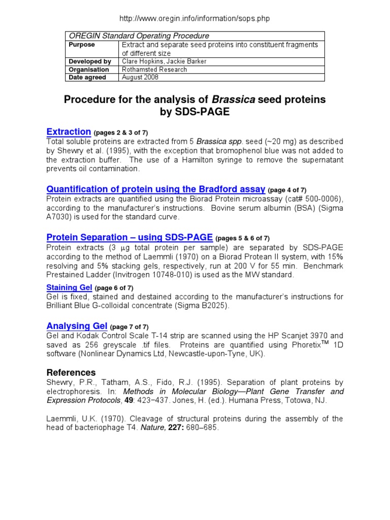
At the end of this lab, students will be able to: • discuss the principles that govern protein separation on discontinuous SDS- PAGE gels. • cast and run SDS-PAGE gels. • analyze the pattern of bands on a stained SDS-PAGE gel • estimate the molecular weight of a protein from its migration on SDS-PAGE gels This lab will introduce you to SDS-PAGE, a simple and Protein analysis by poly acrylamide gel electrophoresis (SDS-PAGE) The protein patterns were analyzed using SDS-PAGE according to Laemmli, (1970) in the first dimension. SDS-PAGE solutions Stock (1) Acrylamide (30 grams) and 0.8 gram bis N.N. methylene bisacrylamide were dissolved in 100 ml distilled water (each component dissolved separately).
Download SDS-PAGE protocol as a PDF . SDS-PAGE, with full name of sodium dodecyl sulfate polyacrylamide gel electrophores, is the most widely used technique to separate proteins from complicated samples of mixture, plays key roles in molecular biology and wide range of subfield of biological research. Being present a electricity, proteins migerate towards the negative … In order to identify proteins by size, protein standards of a known size are loaded along with samples and run under the same conditions. This video presents an introduction to SDS-PAGE by first explaining the theory behind it and later demonstrating its step-by-step procedure.
UIC REERENCE Pub No MAN0007891 Rev C.0 NuPAGE™ Bis-Tris Mini Gels Choosing a well format Thicker 1.5 mm gels with fewer wells are recommended for large samples (>30 μL). Thinner 1 mm gels are recommended for blotting because of better protein transfer. Native SDS-PAGE of LLC-PK 1 cell proteome and model metalloproteins. Neither SDS-PAGE nor BN-PAGE was able to accomplish the goal of acceptable protein separation with retention of enzymatic activity. A new method or effective changes to these protocols were needed to achieve both. We chose to modify SDS-PAGE.
Nov 17, 2015 · Smaller protein-SDS complexes migrate more quickly than larger protein SDS complexes. Within a certain range determined by the porosity of the gel, the migration rate of a protein in the running gel is inversely proportional to the logarithm of its MW. Posted in Protocol Tagged Protocol of SDS-PAGE, Resolving gel buffer, SDS-PAGE, Stacking SDS/PAGE MINI PROTEIN GEL Polyacrylamide gel electrophoresis (PAGE) is a widely used technique for separating proteins. The most widely used method was developed by Laemmli (Nature 227: 690-685, 1970) using the denaturing (SDS) discontinuous method. This protocol relies on the presence
Bio 6 – SDS-PAGE Lab Objectives Upon completion of this laboratory you will understand how to load and run protein samples on an SDS-polyacrylamide gel, stain the gel, and analyze the resulting bands of protein on the gel to estimate the molecular weight of each protein. Peccoud Lab Protocol: visualizing proteins via sodium dodecyl sulfate-polyacrylamide electrophoresis (SDS-PAGE) Introduction. Because cellular extracts contain thousands of different proteins at a wide range of concentrations, it is often difficult to detect and measure specific proteins in these mixes, even when proteins are expressed at high concentrations.
Protein analysis by poly acrylamide gel electrophoresis (SDS-PAGE) The protein patterns were analyzed using SDS-PAGE according to Laemmli, (1970) in the first dimension. SDS-PAGE solutions Stock (1) Acrylamide (30 grams) and 0.8 gram bis N.N. methylene bisacrylamide were dissolved in 100 ml distilled water (each component dissolved separately). INTRODUCTION TO THE PROTEIN ELECTROPHORESIS KIT The Protein Electrophoresis Kit from the Fralin Biotechnology Center contains all the materials needed to prepare samples, run SDS-PAGE gels, visualize the proteins on the gels, and dry the gels to preserve them. The borrower must provide the sample materials (fish, seafood, meat, etc.).
Together, Laemmli–SDS-PAGE and Tricine–SDS-PAGE cover the protein mass range 1–500 kDa. Answering the following questions should help you to identify the optimal solution for a specific sep-aration problem and help you to decide whether this … pdf. PROTOCOL Tricine–SDS-PAGE. Hoi Suparat. Here I describe a protocol for Tricine–SDS-PAGE, which includes efficient methods for Coomassie blue or silver staining and electroblotting, thereby increasing the versatility of the approach. and generates SDS-protein complexes that are mostly characterized by a uniform charge-to-mass
Oct 20, 2018 · The method and detailed procedure of SDS-PAGE for silk proteins are exactly the same as for other proteins, but the electrophoresis profile of silk protein is often unsatisfactory. The main reason is that their molecular masses are too large, and the regenerated liquid silk is easily coagulated and denatured, resulting in a significant adverse effect on normal … SDS/PAGE MINI PROTEIN GEL Polyacrylamide gel electrophoresis (PAGE) is a widely used technique for separating proteins. The most widely used method was developed by Laemmli (Nature 227: 690-685, 1970) using the denaturing (SDS) discontinuous method. This protocol relies on the presence
Use PAGE gels, a Bradford assay, a Lowry assay or a BCA assay. Bovine serum albumin (BSA) is a frequently-used protein standard. Preparation of samples for loading into gels. To denature, use a loading buffer with the anionic denaturing detergent sodium dodecyl sulfate (SDS), and boil the mixture at 95-100°C for 5 minutes. Electrophoresis. 1). UIC REERENCE Pub No MAN0007891 Rev C.0 NuPAGE™ Bis-Tris Mini Gels Choosing a well format Thicker 1.5 mm gels with fewer wells are recommended for large samples (>30 μL). Thinner 1 mm gels are recommended for blotting because of better protein transfer.
Peccoud Lab Protocol: visualizing proteins via sodium dodecyl sulfate-polyacrylamide electrophoresis (SDS-PAGE) Introduction. Because cellular extracts contain thousands of different proteins at a wide range of concentrations, it is often difficult to detect and measure specific proteins in these mixes, even when proteins are expressed at high concentrations. Recommended SDS PAGE Stain Protocols Kits like GelCode Blue from Pierce and Biosafe Coomassie from Biorad are NOT compatible for in-gel digestion and mass spectrometry analysis unless you do a fixing step first.
NuPAGE Bis-Tris Mini Gels Thermo Fisher Scientific. Download SDS-PAGE protocol as a PDF . SDS-PAGE, with full name of sodium dodecyl sulfate polyacrylamide gel electrophores, is the most widely used technique to separate proteins from complicated samples of mixture, plays key roles in molecular biology and wide range of subfield of biological research. Being present a electricity, proteins migerate towards the negative …, SDS-PAGE for protein electrophoresis Aim This section is taked and modified from the protocol provided by Kristian Dreij, Hanna Karlsson in SDS-PAGEprotocolbyAssay-protocol.com. The protocol written by Kristian Dreij, Hanna Karlsson in the course "Applications of Methods in.
(PDF) Optimization of an efficient SDS-PAGE protocol for

SDS-PAGE PROTOCOL NCBS MS-FACILITY. Use PAGE gels, a Bradford assay, a Lowry assay or a BCA assay. Bovine serum albumin (BSA) is a frequently-used protein standard. Preparation of samples for loading into gels. To denature, use a loading buffer with the anionic denaturing detergent sodium dodecyl sulfate (SDS), and boil the mixture at 95-100°C for 5 minutes. Electrophoresis. 1)., SODIUM DODECYL SULFATE-POLYACRYLAMIDE GEL ELECTROPHORESIS (SDS-PAGE) All Hycult Biotech products are subject to strict quality control procedures..
Protein analysis by poly acrylamide gel electrophoresis

SODIUM DODECYL SULFATE-POLYACRYLAMIDE GEL. Introduction to SDS-PAGE. This material is accompanied by a presentation on protein structure and principles behind denaturing samples and discontinuous gel electrophoresis.. The separation of macromolecules in an electric field is called electrophoresis.A very common method for separating proteins by electrophoresis uses a discontinuous polyacrylamide gel as a support … https://fr.wikipedia.org/wiki/Bleu_de_Coomassie Native SDS-PAGE of LLC-PK 1 cell proteome and model metalloproteins. Neither SDS-PAGE nor BN-PAGE was able to accomplish the goal of acceptable protein separation with retention of enzymatic activity. A new method or effective changes to these protocols were needed to achieve both. We chose to modify SDS-PAGE..

SODIUM DODECYL SULFATE-POLYACRYLAMIDE GEL ELECTROPHORESIS (SDS-PAGE) All Hycult Biotech products are subject to strict quality control procedures. Page 3 of 4 Title: SDS-PAGE SOP 8.4. Load Samples. 8.4.1. Using a micropipettor and disposable tips, load 10µL of the Molecular Weight Marker into one well and up to 50µL of each sample into separate wells. 8.4.1.1.Avoid loading samples symmetrically. 8.4.2.
INTRODUCTION TO THE PROTEIN ELECTROPHORESIS KIT The Protein Electrophoresis Kit from the Fralin Biotechnology Center contains all the materials needed to prepare samples, run SDS-PAGE gels, visualize the proteins on the gels, and dry the gels to preserve them. The borrower must provide the sample materials (fish, seafood, meat, etc.). 2-D electrophoresis results. The 2-D protocols described herein are performed using Amersham Biosciences products. Equipment choices are discussed on page 12 and illustrated in Table 1. Introduction to two-dimensional (2-D) electrophoresis Two-dimensional electrophoresis (2-D electrophoresis) is a powerful and widely used
Oct 20, 2018 · The method and detailed procedure of SDS-PAGE for silk proteins are exactly the same as for other proteins, but the electrophoresis profile of silk protein is often unsatisfactory. The main reason is that their molecular masses are too large, and the regenerated liquid silk is easily coagulated and denatured, resulting in a significant adverse effect on normal … Protein gel electrophoresis technical handbook Electrophoresis run conditions 2 For ordering information refer to page XX. For quick reference on the protocol please refer to page XX. 3 ntse Cnto Electrophoresis overview 4 Select precast gel Gel selection guide 8 Gels 10 The resultant SDS-protein complexes are highly
Introduction to SDS-PAGE. This material is accompanied by a presentation on protein structure and principles behind denaturing samples and discontinuous gel electrophoresis.. The separation of macromolecules in an electric field is called electrophoresis.A very common method for separating proteins by electrophoresis uses a discontinuous polyacrylamide gel as a support … interaction permits boiling the protein complex in SDS sample buffer prior to SDS-PAGE analysis. Additional Resources for the HaloTag®Technology Technical Bulletins and Manuals TM260 HaloTag®Technology: Focus on Imaging Technical Manual Promega Publications Efficient high-throughput protein purification using the Magne™ HaloTag®Beads
INTRODUCTION TO THE PROTEIN ELECTROPHORESIS KIT The Protein Electrophoresis Kit from the Fralin Biotechnology Center contains all the materials needed to prepare samples, run SDS-PAGE gels, visualize the proteins on the gels, and dry the gels to preserve them. The borrower must provide the sample materials (fish, seafood, meat, etc.). Sodium dodecyl sulfate (SDS)-polyacrylamide gel electrophoresis (PAGE) is an analytical method that enables protein separation based on their molecular mass. SDS is an anionic detergent, which facilitates the denaturation of the native proteins by disturbing the non-covalent forces.
— If protein phosphorylation is to be studied, include phosphatase inhibitors such as okadaic acid, calyculin A, and vanadate. When working with a new sample, use at least two different cell disruption protocols and compare the protein yield (by protein assay) and qualitative protein content (by SDS-PAGE) Bio 6 – SDS-PAGE Lab Objectives Upon completion of this laboratory you will understand how to load and run protein samples on an SDS-polyacrylamide gel, stain the gel, and analyze the resulting bands of protein on the gel to estimate the molecular weight of each protein.
Use PAGE gels, a Bradford assay, a Lowry assay or a BCA assay. Bovine serum albumin (BSA) is a frequently-used protein standard. Preparation of samples for loading into gels. To denature, use a loading buffer with the anionic denaturing detergent sodium dodecyl sulfate (SDS), and boil the mixture at 95-100°C for 5 minutes. Electrophoresis. 1). Oct 20, 2018 · SDS-PAGE (Polyacrylamide Gel Electrophoresis), is an analytical method used to separate components of a protein mixture based on their size. The technique is based upon the principle that a charged molecule will migrate in an electric field …
Protein Isolation Protocols › Protein Purification Protocols › SDS PAGE Protocols BenchMark™ Pre-Stained Protein Ladder One-Dimensional SDS Gel Electrophoresis of Peptides and Small Proteins with Pre-Cast Gels — If protein phosphorylation is to be studied, include phosphatase inhibitors such as okadaic acid, calyculin A, and vanadate. When working with a new sample, use at least two different cell disruption protocols and compare the protein yield (by protein assay) and qualitative protein content (by SDS-PAGE)
SODIUM DODECYL SULFATE-POLYACRYLAMIDE GEL ELECTROPHORESIS (SDS-PAGE) All Hycult Biotech products are subject to strict quality control procedures. Introduction to SDS-PAGE. This material is accompanied by a presentation on protein structure and principles behind denaturing samples and discontinuous gel electrophoresis.. The separation of macromolecules in an electric field is called electrophoresis.A very common method for separating proteins by electrophoresis uses a discontinuous polyacrylamide gel as a support …
Adapted from protocol accompanying Hybond ECL Membrane Materials Transfer Buffer 1x SDS Running Buffer in 20% Methanol 1x PBS/0.1% Tween 20 Blotting buffer, store at 4 ºC 5% milk in 1x PBS/0.1% Tween 20 Protocol 1. Run SDS-PAGE. 2. Wet membrane in H2O. Soak membrane in transfer buffer for 10 min. 3. In SDS- PAGE, the protein mixture is denatured by heating at 100 qC in the presence of excess SDS and a reducing reagent is employed to break disu lfide bonds. Under these conditions, all reduced polypeptide bind the same amount of SDS on a weight basis (1.4g SDS/g polypeptide) independent of the amino acid composition and sequence of the protein.

In SDS- PAGE, the protein mixture is denatured by heating at 100 qC in the presence of excess SDS and a reducing reagent is employed to break disu lfide bonds. Under these conditions, all reduced polypeptide bind the same amount of SDS on a weight basis (1.4g SDS/g polypeptide) independent of the amino acid composition and sequence of the protein. (Denaturing) SDS Page Protein Gel Protocol. Solutions . 1. 30% acrylamide/.8%bisacrylamide 30g Acrylamide (store in brown light sensitive container) .8g Bisacrylamide