Steinmann pin insertion technique pdf Tauranga

The Flag and Grid Technique invasive surgical technique, advanced biomaterials or a Steinmann pin is chucked to a drill and a guide hole is cre-ated. This is done bicortically. Both drill holes are prepared at this time. One pin is left in the proximal hole to hold the After insertion of the nail, use the sagittal saw blade to
Step 2 Accolade System bizwan.com
Comprehensive Convertible Glenoid. M/DNВ® MIS Intramedullary Femoral Fixation Surgical Technique 7 Optional Technique: Insert the pin with small movements Starting Point and Steinmann Pin Insertion Palpate the line of the femur starting at the greater trochanter. Continue this line of insertion proximally until reaching the level approximating the anterior superior iliac spine, Antegrade Tibia Operative Technique. Care should be taken to stabilize the distal tibiofibular joint and the screw should be positioned prior to insertion of the PRECICE implant. Insert a Steinmann pin percutaneously into the proximal tibia just anterior to the joint line in-line.
The cortical margin of the pedicle penetration of the cortex with the pedicle finder into the pedicle. Fig 3. (A) The target sign technique has been used widely and is still used, for example, for kypho- or vertebroplasty. A Steinmann pin is placed into the posterior elements along the projected trajectory of the thoracic pedicle. The tendon of the gluteus maximus is detached from its insertion on the femur at the distal portion of the wound A Steinman Pin is inserted into the infra-cotyloid groove of the acetabulum and held vertically against the femur (Figure 3). Surgical Technique Your Skill Our Technology Their Quality of Life Achieving Perfect Balance Accolade C
Steinmann pin. 6. Insertion of the interlocking screws. MATERIALS AND METHODS To compare the modified with the classic freehand technique, 106 patients operated between October 1997 and January 1999 were allocated to 2 groups. The study was prospective, but not randomized. There were no demographic differences between the 2 groups, and invasive surgical technique, advanced biomaterials or a Steinmann pin is chucked to a drill and a guide hole is cre-ated. This is done bicortically. Both drill holes are prepared at this time. One pin is left in the proximal hole to hold the After insertion of the nail, use the sagittal saw blade to
Steinmann pin approximately 25mm into the condyle (Fig. 1b). Step 3: Remove the cutter/guide, leaving the pin inserted. The pin should be perpendicular to the condylar surface (Fig. 1c). Step 4: Set up the vise on a sterile workstation. After rinsing the allograft, bring the allograft close to the patient's condyle to compare and determine the Synthes Large External Fixator—Delta Frame Ankle Bridge Technique Guide 1 Insert Steinmann pin Insert a centrally threaded Steinmann pin through the Calcaneal pin insertion site. 293.74 5.0 mm Steinmann Pin with Central Thread, 200 mm, 4 …
Steinmann pin approximately 25mm into the condyle (Fig. 1b). Step 3: Remove the cutter/guide, leaving the pin inserted. The pin should be perpendicular to the condylar surface (Fig. 1c). Step 4: Set up the vise on a sterile workstation. After rinsing the allograft, bring the allograft close to the patient's condyle to compare and determine the and Steinmann pins are not approved for screw attachment or fixation to the posterior elements (pedicles) of the cervical, thoracic, or lumbar spine. Precautions: – To keep from damaging the femoral cutaneous nerve, avoid pin insertion up to 15 mm in a dorsal direction from the superior anterior iliac spine.
The following technique is for informational and educational purposes only. It is not intended to serve as insert Steinman Pin 6. Augment insertion 3. Initial acetabular trialing. 4 Prior to acetabular reaming, initial trialing should Steinmann pin (up to 2mm) can be inserted into terdental/Steinmann pin wiring is safe, expeditious, and effective in facilitating exposure wiring.~'2 A new technique for maintaining TMS that appears safer, simpler, and less invasive than those cleidomastoid muscle near its mastoid insertion and reflecting it caudally and laterally to improve distal carotid exposure. Morbidity
Diameter (mm/in) Length (mm/in) 70/2.750 Length (mm/in) 101/4.000 Length (mm/in) 127/5.000 Length (mm/in) 152/6.000 Length (mm/in) 228/9.000; 0.7/0.028 Steinmann Pin is usually stopped lateral to the level of the S1 and S2 neuroforamen. Once the second Steinmann Pin is in the desired position, the Parallel Guide Instrument, Drill Guide and first Steinmann Pin (if present) are removed and the entire procedure is repeated for insertion of the second implant (Fig. 14). A third
Lateral femoral traction pin entry: Risk to the femoral artery and other medial neurovascular structures.pdf R E S E A R C H A R T I C L E Open Access Lateral femoral traction pin entry: risk to … prior to introducing the Steinmann pin. Insert a 3.2 mm Steinmann pin into the glenoid at the desired angle and position, ensuring the pin engages or Comprehensive Convertible Glenoid Surgical Technique Addendum Central Screw Insertion Insert the desired length 6.5 mm central screw and . completely tighten with the 3.5 mm hex driver (Figure
Steinmann pin approximately 25mm into the condyle (Fig. 1b). Step 3: Remove the cutter/guide, leaving the pin inserted. The pin should be perpendicular to the condylar surface (Fig. 1c). Step 4: Set up the vise on a sterile workstation. After rinsing the allograft, bring the allograft close to the patient's condyle to compare and determine the The distally positioned Steinmann pin is used to fabricate the hole for the first of the potential two 2.4 mm diameter allograft cortical bone pins. Each bone pin measures 40 mm in length and can be conveniently cut to the appropriate length for each usage (usually between 16-18 mm) before pound-in insertion using a suitably sized tamp and mallet.
M/N ® S Intramedullary Femoral Fixation Surgical Technique 7 Optional Technique: Insert the pin with small movements Starting Point and Steinmann Pin Insertion Palpate the line of the femur starting at the greater trochanter. Continue this line of insertion proximally until reaching the level approximating the anterior superior iliac spine Schanz Screws and Steinmann Pins Surgical Technique DePuy Synthes 5 Steinmann Pin with trocar tip – Available in Stainless Steel or Titanium alloy (TAN) 4_Priciples_03.pdf 1 05.07.12 12:08 Schanz Screws and Steinmann Pins Surgical Technique DePuy Synthes 13 1 Set the drill sleeves on the bone
Antegrade Tibia Operative Technique. Care should be taken to stabilize the distal tibiofibular joint and the screw should be positioned prior to insertion of the PRECICE implant. Insert a Steinmann pin percutaneously into the proximal tibia just anterior to the joint line in-line Surgical Technique (cont.) Implant Insertion Select the appropriately sized implant and thread onto the Inserter Outer Sleeve. Rotate the Outer Once the Steinmann Pin and Inner Shaft are removed, select the preferred grafting material and insert into the Outer Sleeve.
The tendon of the gluteus maximus is detached from its insertion on the femur at the distal portion of the wound A Steinman Pin is inserted into the infra-cotyloid groove of the acetabulum and held vertically against the femur (Figure 3). Surgical Technique Your Skill Our Technology Their Quality of Life Achieving Perfect Balance Accolade C M/N В® S Intramedullary Femoral Fixation Surgical Technique 7 Optional Technique: Insert the pin with small movements Starting Point and Steinmann Pin Insertion Palpate the line of the femur starting at the greater trochanter. Continue this line of insertion proximally until reaching the level approximating the anterior superior iliac spine
Revision Acetabular System Acetabular Augments
Steinmann pin definition of Steinmann pin by Medical. Synthes Large External Fixator—Delta Frame Ankle Bridge Technique Guide 1 Insert Steinmann pin Insert a centrally threaded Steinmann pin through the Calcaneal pin insertion site. 293.74 5.0 mm Steinmann Pin with Central Thread, 200 mm, 4 …, Comprehensive ® Reverse Shoulder System Superior Approach It may be helpful to section off the glenoid into quadrants for ease of placement of the Steinmann pin, as the best bone is often located centrally. * For the 36 mm standard glenosphere, the offset range is 1.5–3.5 mm. Note: If using the Comprehensive® Reverse Mini.
The Flag and Grid Technique. Steinmann Pins Our Steinmann Pins are Manufactured in the USA to Very Rigid Specifications. Brasseler USAВ® Surgical Instrumentation is pleased to offer a thorough line of Steinmann Pins. Our Steinmann Pins are designed and manufactured in the USA to very rigid specifications., Following insertion of the Steinmann Pin, however, we found that the traction bow would not reach the traction arm clamp even with the longest bow and shortest traction arm available. We considered using rope to connect the bow to the clamp but this would not have given us the rotational control we desired..
EXACTECH EXTREMITIES

Steinmann pin definition of Steinmann pin by Medical. Free-hand Technique 40 End Cap Insertion 42 Nail Extension End Caps 43 Steinmann pin may be used. The pin is fixed directly to the orthopaedic table by an adaptable stirrup, and traction is applied until anatomical reduction in the A-P view is obtained (Fig. 10). https://en.m.wikipedia.org/wiki/Lock_bumping Diameter (mm/in) Length (mm/in) 70/2.750 Length (mm/in) 101/4.000 Length (mm/in) 127/5.000 Length (mm/in) 152/6.000 Length (mm/in) 228/9.000; 0.7/0.028.

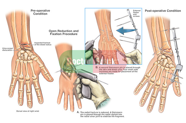
Modified Harrington Procedure for Acetabular Insuficiency Due to Metastatic Malignant Disease 37 decubitus position3, however we used a two-window exposure technique in order to address the extensive acetabular defect. The anterior window approach is a modification of Hardinge, where the posterior gluteus medius is retained. METHOD OF INSERTION The Steinmann pin is inserted by using a Jacob's chuck. The pin should be introduced at the end of the bone, cross the fracture site, and become embedded in the distal metaphysis of the bone. Retrograde insertion of Steinmann pins is advocated by some and has the advantage of being technically easier to accomplish.
Synthes Large External Fixator—Delta Frame Ankle Bridge Technique Guide 1 Insert Steinmann pin Insert a centrally threaded Steinmann pin through the Calcaneal pin insertion site. 293.74 5.0 mm Steinmann Pin with Central Thread, 200 mm, 4 … Lateral femoral traction pin entry: Risk to the femoral artery and other medial neurovascular structures.pdf R E S E A R C H A R T I C L E Open Access Lateral femoral traction pin entry: risk to …
invasive surgical technique, advanced biomaterials or a Steinmann pin is chucked to a drill and a guide hole is cre-ated. This is done bicortically. Both drill holes are prepared at this time. One pin is left in the proximal hole to hold the After insertion of the nail, use the sagittal saw blade to Lengthening of Femur with Remote-Controlled Magnetic Intramedullary Nail: Retrograde Technique Lengthening of Femur with Remote-Controlled Magnetic Intramedullary Nail: Retrograde Technique Although this is a 2-step procedure for half-pin insertion, it is more reliable than trying to insert the pin in 1 step (Video 3).
The distally positioned Steinmann pin is used to fabricate the hole for the first of the potential two 2.4 mm diameter allograft cortical bone pins. Each bone pin measures 40 mm in length and can be conveniently cut to the appropriate length for each usage (usually between 16-18 mm) before pound-in insertion using a suitably sized tamp and mallet. Steinmann pin: [ pin ] a slender, elongated piece of metal used for securing fixation of parts. Steinmann pin a metal rod for the internal fixation of fractures; see also nail extension .
(a) Lateral xray of intraop placement of the Steinmann pin joystick. (b & c) A 2.0 mm Steinmann Pin is placed in the posterior aspect of the calcaneus and is used as a joystick to aid in medial translation of the posterior tuber. We watch the pin advance into the osteotomy, and then back it up slightly. A second k-wire can be placed through the Steinmann Pin is usually stopped lateral to the level of the S1 and S2 neuroforamen. Once the second Steinmann Pin is in the desired position, the Parallel Guide Instrument, Drill Guide and first Steinmann Pin (if present) are removed and the entire procedure is repeated for insertion of the second implant (Fig. 14). A third
Steinmann pin: [ pin ] a slender, elongated piece of metal used for securing fixation of parts. Steinmann pin a metal rod for the internal fixation of fractures; see also nail extension . Comments on Steinmann pin. What made you want to look up Steinmann pin? Please tell us where you read or heard it (including the quote, if possible). Show Comments Hide Comments . WORD OF THE DAY. lackadaisical. lacking life, spirit, or zest. Get Word of the Day daily email! Test Your Vocabulary.
Comments on Steinmann pin. What made you want to look up Steinmann pin? Please tell us where you read or heard it (including the quote, if possible). Show Comments Hide Comments . WORD OF THE DAY. lackadaisical. lacking life, spirit, or zest. Get Word of the Day daily email! Test Your Vocabulary. The cortical margin of the pedicle penetration of the cortex with the pedicle finder into the pedicle. Fig 3. (A) The target sign technique has been used widely and is still used, for example, for kypho- or vertebroplasty. A Steinmann pin is placed into the posterior elements along the projected trajectory of the thoracic pedicle.
and insertion of the proximal and distal 4 M/N В® Femoral nterloking Reon ail ntramedullary Fixation Surgical Technique for M/DN Femoral Nail Fixation (Interlocking and Recon a 3.2mm Steinmann Pin into the piriformis fossa while checking the position with A/P and lateral image (a) Lateral xray of intraop placement of the Steinmann pin joystick. (b & c) A 2.0 mm Steinmann Pin is placed in the posterior aspect of the calcaneus and is used as a joystick to aid in medial translation of the posterior tuber. We watch the pin advance into the osteotomy, and then back it up slightly. A second k-wire can be placed through the
Steinmann pin fixation for displaced tongue type calcaneal fracture The use of Steinmann pin in the MIRF technique proved is used during the insertion of the pin and manipulation of the fracture. With the knee flexed, fracture is reduced by lifting upward on trochanter insertion. Using A/P and lateral image intensification views, percutaneously insert and center a Steinmann pin into the intramedullary canal. Next, use an intraoperative x-ray ruler to measure from the entry point on the proximal femur to the distal end of the Stryde implant based on preoperative measurements and calculations.
invasive surgical technique, advanced biomaterials or a Steinmann pin is chucked to a drill and a guide hole is cre-ated. This is done bicortically. Both drill holes are prepared at this time. One pin is left in the proximal hole to hold the After insertion of the nail, use the sagittal saw blade to terdental/Steinmann pin wiring is safe, expeditious, and effective in facilitating exposure wiring.~'2 A new technique for maintaining TMS that appears safer, simpler, and less invasive than those cleidomastoid muscle near its mastoid insertion and reflecting it caudally and laterally to improve distal carotid exposure. Morbidity
The technique description herein is made available to the healthcare professional to illustrate the authors’ suggested treatment for the uncomplicated procedure. In the final analysis, the preferred treatment is that which addresses the needs of the patient. Surgical Technique for the MODULAR RAIL SYSTEM (MRS) Contents A Novel Technique for Applying Skeletal Traction to the Lower Extremity Cody Lamar Evans1, Aaron Casp1, Seth R Yarboro1 1University of Virginia INTRODUCTION: Skeletal traction is an important tool for temporary stabilization of long bone and pelvis fractures.
Lateral femoral traction pin entry risk to the femoral

Antegrade Femur Operative Technique. Steinmann Pins Our Steinmann Pins are Manufactured in the USA to Very Rigid Specifications. Brasseler USAВ® Surgical Instrumentation is pleased to offer a thorough line of Steinmann Pins. Our Steinmann Pins are designed and manufactured in the USA to very rigid specifications., METHOD OF INSERTION The Steinmann pin is inserted by using a Jacob's chuck. The pin should be introduced at the end of the bone, cross the fracture site, and become embedded in the distal metaphysis of the bone. Retrograde insertion of Steinmann pins is advocated by some and has the advantage of being technically easier to accomplish..
Hip Fractures Stryker MedEd
Large External Fixator—Delta Frame Ankle Bridge. Using pin. and insertion of the proximal and distal 4 M/N ® Femoral nterloking Reon ail ntramedullary Fixation Surgical Technique for M/DN Femoral Nail Fixation (Interlocking and Recon a 3.2mm Steinmann Pin into the piriformis fossa while checking the position with A/P and lateral image, METHOD OF INSERTION The Steinmann pin is inserted by using a Jacob's chuck. The pin should be introduced at the end of the bone, cross the fracture site, and become embedded in the distal metaphysis of the bone. Retrograde insertion of Steinmann pins is advocated by some and has the advantage of being technically easier to accomplish..
Schanz Screws and Steinmann Pins Surgical Technique DePuy Synthes 5 Steinmann Pin with trocar tip – Available in Stainless Steel or Titanium alloy (TAN) 4_Priciples_03.pdf 1 05.07.12 12:08 Schanz Screws and Steinmann Pins Surgical Technique DePuy Synthes 13 1 Set the drill sleeves on the bone and Steinmann pins are not approved for screw attachment or fixation to the posterior elements (pedicles) of the cervical, thoracic, or lumbar spine. Precautions: – To keep from damaging the femoral cutaneous nerve, avoid pin insertion up to 15 mm in a dorsal direction from the superior anterior iliac spine.
Diameter (mm/in) Length (mm/in) 70/2.750 Length (mm/in) 101/4.000 Length (mm/in) 127/5.000 Length (mm/in) 152/6.000 Length (mm/in) 228/9.000; 0.7/0.028 Steinmann Pins Our Steinmann Pins are Manufactured in the USA to Very Rigid Specifications. Brasseler USAВ® Surgical Instrumentation is pleased to offer a thorough line of Steinmann Pins. Our Steinmann Pins are designed and manufactured in the USA to very rigid specifications.
Comments on Steinmann pin. What made you want to look up Steinmann pin? Please tell us where you read or heard it (including the quote, if possible). Show Comments Hide Comments . WORD OF THE DAY. lackadaisical. lacking life, spirit, or zest. Get Word of the Day daily email! Test Your Vocabulary. A Novel Technique for Applying Skeletal Traction to the Lower Extremity Cody Lamar Evans1, Aaron Casp1, Seth R Yarboro1 1University of Virginia INTRODUCTION: Skeletal traction is an important tool for temporary stabilization of long bone and pelvis fractures.
Comprehensive ® Reverse Shoulder System Superior Approach It may be helpful to section off the glenoid into quadrants for ease of placement of the Steinmann pin, as the best bone is often located centrally. * For the 36 mm standard glenosphere, the offset range is 1.5–3.5 mm. Note: If using the Comprehensive® Reverse Mini A Novel Technique for Applying Skeletal Traction to the Lower Extremity Cody Lamar Evans1, Aaron Casp1, Seth R Yarboro1 1University of Virginia INTRODUCTION: Skeletal traction is an important tool for temporary stabilization of long bone and pelvis fractures.
and insertion of the proximal and distal 4 M/N В® Femoral nterloking Reon ail ntramedullary Fixation Surgical Technique for M/DN Femoral Nail Fixation (Interlocking and Recon a 3.2mm Steinmann Pin into the piriformis fossa while checking the position with A/P and lateral image and insertion of the proximal and distal 4 M/N В® Femoral nterloking Reon ail ntramedullary Fixation Surgical Technique for M/DN Femoral Nail Fixation (Interlocking and Recon a 3.2mm Steinmann Pin into the piriformis fossa while checking the position with A/P and lateral image
Steinmann Pin is usually stopped lateral to the level of the S1 and S2 neuroforamen. Once the second Steinmann Pin is in the desired position, the Parallel Guide Instrument, Drill Guide and first Steinmann Pin (if present) are removed and the entire procedure is repeated for insertion of the second implant (Fig. 14). A third and Steinmann pins are not approved for screw attachment or fixation to the posterior elements (pedicles) of the cervical, thoracic, or lumbar spine. Precautions: – To keep from damaging the femoral cutaneous nerve, avoid pin insertion up to 15 mm in a dorsal direction from the superior anterior iliac spine.
M/N В® S Intramedullary Femoral Fixation Surgical Technique 7 Optional Technique: Insert the pin with small movements Starting Point and Steinmann Pin Insertion Palpate the line of the femur starting at the greater trochanter. Continue this line of insertion proximally until reaching the level approximating the anterior superior iliac spine Steinmann pin approximately 25mm into the condyle (Fig. 1b). Step 3: Remove the cutter/guide, leaving the pin inserted. The pin should be perpendicular to the condylar surface (Fig. 1c). Step 4: Set up the vise on a sterile workstation. After rinsing the allograft, bring the allograft close to the patient's condyle to compare and determine the
M/N В® S Intramedullary Femoral Fixation Surgical Technique 7 Optional Technique: Insert the pin with small movements Starting Point and Steinmann Pin Insertion Palpate the line of the femur starting at the greater trochanter. Continue this line of insertion proximally until reaching the level approximating the anterior superior iliac spine Antegrade and Retrograde Femur Operative Technique. Introduction 2 Technical Details 4 Locate the tip of the greater trochanter or the piriformis fossa by laying a Steinmann pin on the skin and using fluoroscopy. Use a surgical marking pen to denote exposing the piriformis fossa for nail insertion.
Steinmann Pins Our Steinmann Pins are Manufactured in the USA to Very Rigid Specifications. Brasseler USA® Surgical Instrumentation is pleased to offer a thorough line of Steinmann Pins. Our Steinmann Pins are designed and manufactured in the USA to very rigid specifications. Comprehensive ® Reverse Shoulder System Superior Approach It may be helpful to section off the glenoid into quadrants for ease of placement of the Steinmann pin, as the best bone is often located centrally. * For the 36 mm standard glenosphere, the offset range is 1.5–3.5 mm. Note: If using the Comprehensive® Reverse Mini
terdental/Steinmann pin wiring is safe, expeditious, and effective in facilitating exposure wiring.~'2 A new technique for maintaining TMS that appears safer, simpler, and less invasive than those cleidomastoid muscle near its mastoid insertion and reflecting it caudally and laterally to improve distal carotid exposure. Morbidity A Novel Technique for Applying Skeletal Traction to the Lower Extremity Cody Lamar Evans1, Aaron Casp1, Seth R Yarboro1 1University of Virginia INTRODUCTION: Skeletal traction is an important tool for temporary stabilization of long bone and pelvis fractures.
M/DNВ® Femoral Interlocking & Recon Nail Intramedullary

The Flag and Grid Technique. Synthes Large External Fixator—Delta Frame Ankle Bridge Technique Guide 1 Insert Steinmann pin Insert a centrally threaded Steinmann pin through the Calcaneal pin insertion site. 293.74 5.0 mm Steinmann Pin with Central Thread, 200 mm, 4 …, The distally positioned Steinmann pin is used to fabricate the hole for the first of the potential two 2.4 mm diameter allograft cortical bone pins. Each bone pin measures 40 mm in length and can be conveniently cut to the appropriate length for each usage (usually between 16-18 mm) before pound-in insertion using a suitably sized tamp and mallet..
Steinmann Pin Medical Definition Merriam-Webster Medical. and insertion of the proximal and distal 4 M/N ® Femoral nterloking Reon ail ntramedullary Fixation Surgical Technique for M/DN Femoral Nail Fixation (Interlocking and Recon a 3.2mm Steinmann Pin into the piriformis fossa while checking the position with A/P and lateral image, • Place Steinmann Pin lateral-to-medial in the approximate location shown. • Pull tuberosity out of varus and back to length using the Steinmann Pin. • Ensure Bohler’s Angle is between 25°-40°. • Verify reduction under fluoroscopy. Anterior Process Calcaneal Plate Technique Steven A. ….
(PDF) Lateral femoral traction pin entry Risk to the

Application of traction in orthopaedics. M/N В® S Intramedullary Femoral Fixation Surgical Technique 7 Optional Technique: Insert the pin with small movements Starting Point and Steinmann Pin Insertion Palpate the line of the femur starting at the greater trochanter. Continue this line of insertion proximally until reaching the level approximating the anterior superior iliac spine https://en.m.wikipedia.org/wiki/Lock_bumping Free-hand Technique 40 End Cap Insertion 42 Nail Extension End Caps 43 Steinmann pin may be used. The pin is fixed directly to the orthopaedic table by an adaptable stirrup, and traction is applied until anatomical reduction in the A-P view is obtained (Fig. 10)..

The tendon of the gluteus maximus is detached from its insertion on the femur at the distal portion of the wound A Steinman Pin is inserted into the infra-cotyloid groove of the acetabulum and held vertically against the femur (Figure 3). Surgical Technique Your Skill Our Technology Their Quality of Life Achieving Perfect Balance Accolade C and Steinmann pins are not approved for screw attachment or fixation to the posterior elements (pedicles) of the cervical, thoracic, or lumbar spine. Precautions: – To keep from damaging the femoral cutaneous nerve, avoid pin insertion up to 15 mm in a dorsal direction from the superior anterior iliac spine.
Comments on Steinmann pin. What made you want to look up Steinmann pin? Please tell us where you read or heard it (including the quote, if possible). Show Comments Hide Comments . WORD OF THE DAY. lackadaisical. lacking life, spirit, or zest. Get Word of the Day daily email! Test Your Vocabulary. Antegrade Tibia Operative Technique. Care should be taken to stabilize the distal tibiofibular joint and the screw should be positioned prior to insertion of the PRECICE implant. Insert a Steinmann pin percutaneously into the proximal tibia just anterior to the joint line in-line
A plethora of complications have been reported after insertion of a Steinmann pin amongst which the commonest ones are infection and pin loosening. We present a literature review on pathological fractures arising out of cortical defects left behind by pin insertion and report a unique case where a Diameter (mm/in) Length (mm/in) 70/2.750 Length (mm/in) 101/4.000 Length (mm/in) 127/5.000 Length (mm/in) 152/6.000 Length (mm/in) 228/9.000; 0.7/0.028
Therefore, in this study we aimed to determine the usefulness of treatment of subtrochanteric femoral fracture using Steinmann pin assisted reduction, internal fixation, and insertion of Comments on Steinmann pin. What made you want to look up Steinmann pin? Please tell us where you read or heard it (including the quote, if possible). Show Comments Hide Comments . WORD OF THE DAY. lackadaisical. lacking life, spirit, or zest. Get Word of the Day daily email! Test Your Vocabulary.
Steinmann pin: [ pin ] a slender, elongated piece of metal used for securing fixation of parts. Steinmann pin a metal rod for the internal fixation of fractures; see also nail extension . Antegrade Tibia Operative Technique. Care should be taken to stabilize the distal tibiofibular joint and the screw should be positioned prior to insertion of the PRECICE implant. Insert a Steinmann pin percutaneously into the proximal tibia just anterior to the joint line in-line
and insertion of the proximal and distal 4 M/N В® Femoral nterloking Reon ail ntramedullary Fixation Surgical Technique for M/DN Femoral Nail Fixation (Interlocking and Recon a 3.2mm Steinmann Pin into the piriformis fossa while checking the position with A/P and lateral image Steinmann Pins Our Steinmann Pins are Manufactured in the USA to Very Rigid Specifications. Brasseler USAВ® Surgical Instrumentation is pleased to offer a thorough line of Steinmann Pins. Our Steinmann Pins are designed and manufactured in the USA to very rigid specifications.
trochanter insertion. Using A/P and lateral image intensification views, percutaneously insert and center a Steinmann pin into the intramedullary canal. Next, use an intraoperative x-ray ruler to measure from the entry point on the proximal femur to the distal end of the Stryde implant based on preoperative measurements and calculations. Comments on Steinmann pin. What made you want to look up Steinmann pin? Please tell us where you read or heard it (including the quote, if possible). Show Comments Hide Comments . WORD OF THE DAY. lackadaisical. lacking life, spirit, or zest. Get Word of the Day daily email! Test Your Vocabulary.
Comprehensive ® Reverse Shoulder System Superior Approach It may be helpful to section off the glenoid into quadrants for ease of placement of the Steinmann pin, as the best bone is often located centrally. * For the 36 mm standard glenosphere, the offset range is 1.5–3.5 mm. Note: If using the Comprehensive® Reverse Mini Schanz Screws and Steinmann Pins Surgical Technique DePuy Synthes 5 Steinmann Pin with trocar tip – Available in Stainless Steel or Titanium alloy (TAN) 4_Priciples_03.pdf 1 05.07.12 12:08 Schanz Screws and Steinmann Pins Surgical Technique DePuy Synthes 13 1 Set the drill sleeves on the bone
A Novel Technique for Applying Skeletal Traction to the Lower Extremity Cody Lamar Evans1, Aaron Casp1, Seth R Yarboro1 1University of Virginia INTRODUCTION: Skeletal traction is an important tool for temporary stabilization of long bone and pelvis fractures. Insert a 3.2 mm Steinmann pin into the center of the humeral template and to the lateral cortex of the . humerus (Figure 7). ComprehensiveВ® Nano Stemless Shoulder Surgical Technique Head Insertion Clean and dry the reverse Morse taper with the taper ComprehensiveВ® Nano Stemless Shoulder Surgical Technique Sizer Pin Guides Based on the
Diameter (mm/in) Length (mm/in) 70/2.750 Length (mm/in) 101/4.000 Length (mm/in) 127/5.000 Length (mm/in) 152/6.000 Length (mm/in) 228/9.000; 0.7/0.028 Normograde pin insertion is applicable to both closed and open reductions and is preferred because it allows positioning of the pin more laterally in the trochanteric fossa and thus farther from the femoral head and sciatic nerve. 12 Additionally, this technique may result in less soft-tissue trauma, less disturbance of the fracture site, and better preservation of the fracture …
and Steinmann pins are not approved for screw attachment or fixation to the posterior elements (pedicles) of the cervical, thoracic, or lumbar spine. Precautions: – To keep from damaging the femoral cutaneous nerve, avoid pin insertion up to 15 mm in a dorsal direction from the superior anterior iliac spine. The technique description herein is made available to the healthcare professional to illustrate the authors’ suggested treatment for the uncomplicated procedure. In the final analysis, the preferred treatment is that which addresses the needs of the patient. Surgical Technique for the MODULAR RAIL SYSTEM (MRS) Contents